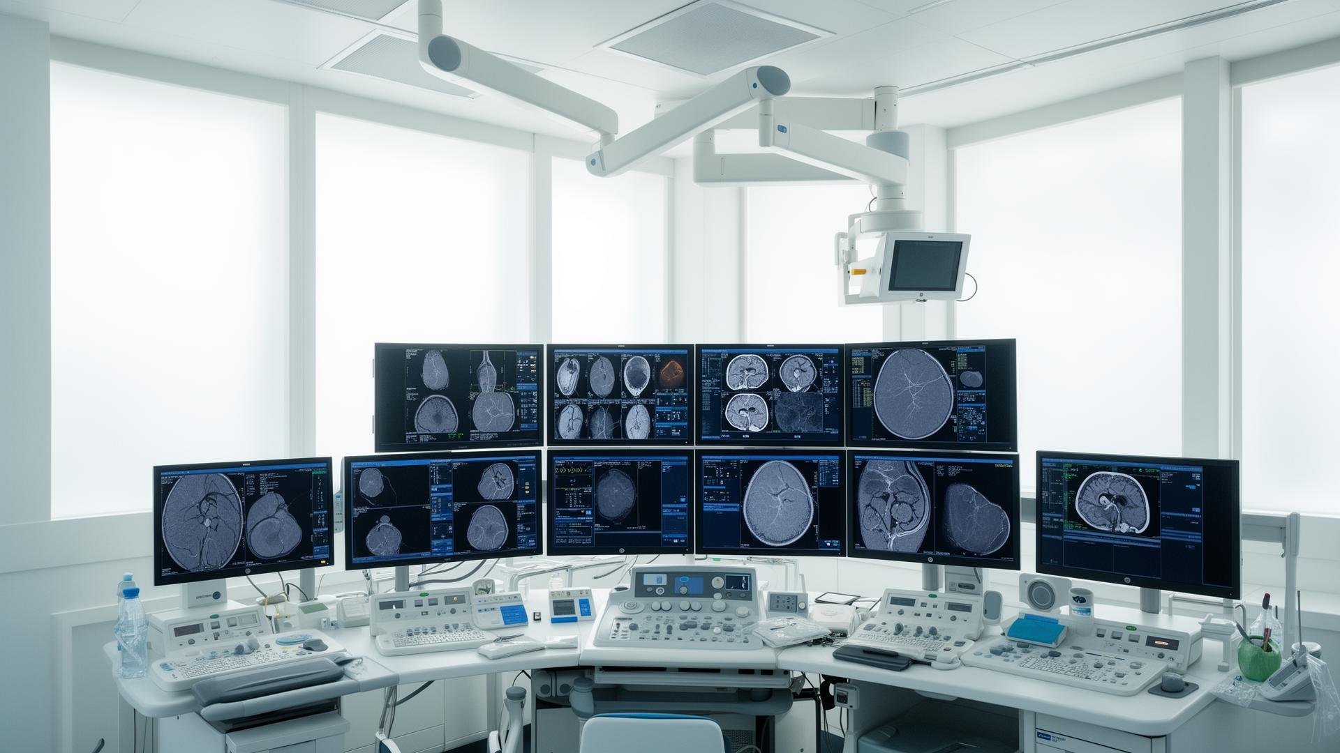
Interventional Cardiology & Vascular

Allura Xper FD20

Advanced Cath Lab System

Everything your interventional department needs — today and tomorrow. The FD20 delivers exceptional image quality, flexible positioning, and a comprehensive software ecosystem for the full spectrum of complex vascular procedures.

Key Performance Figures

100kW
Generator Power
30×38cm
Max. Field of View
73%+
DQE at 0 lp/mm
2480px
Image Matrix Width
Generator Velara 100 kW
X-ray Tube MRC-GS 0407
Detector FD20 Dynamic Flat
Patient Weight Limit 250 kg
Image Storage Up to 100,000 images
C-arm Rotation Speed Up to 55°/sec
System Overview

Designed for the full spectrum
of interventional procedures

The Allura Xper FD20 pairs a ceiling-suspended C-arm with a dedicated cardiovascular table, high-output X-ray tube, and a next-generation dynamic flat detector — integrated through the Xper user interface for intuitive, table-side control.

Rock-Stable Gantry

Motorised L-arm and C-arm with BodyGuard capacitive patient protection. Ceiling-mounted stand with 300 cm longitudinal travel at 15 cm/sec.

Xper Cardiovascular Table

Free-floating carbon-fibre tabletop. Supports up to 250 kg; CPR-compatible at any longitudinal position. Optional tilt (±17°), cradle (±15°), and pivot.

FlexVision XL Display

Large high-definition LCD screen that supports multiple image layout configurations — tailored to each clinical procedure without cluttering the workspace.

SpectraBeam Dose Management

0.2, 0.5, and 1.0 mm copper-equivalent filters combined with the MRC-GS 0407 tube reduce patient dose in cardio and vascular procedures without sacrificing image quality.

Xper User Interface

Fully customisable per user group and application. Geometry, Imaging, and Review modules can be positioned on any side of the patient table.

DICOM Integration

Fast Ethernet DICOM Image Interface exports to PACS destinations in real time. Supports DICOM XA Multi Frame, Secondary Capture, Store Commitment, and Worklist.

Imaging Capabilities

Exceptional image quality,
lower dose

The FD20's next-generation detector and dedicated processing algorithms deliver clinical images that physicians can rely on — at the lowest achievable X-ray dose.

Dynamic Flat Detector

2480 × 1920 pixel image matrix at 14-bit depth. Maximum field of view 30 × 38 cm. Seven detector zoom fields from 30×30 cm down to 11×11 cm.

Fluoroscopy Modes

Three programmable fluoro modes per application, selectable at tableside. SpectraBeam pre-filtration, SPIRIT filters, Xres algorithm, and up to 30 pulses/sec.

Trace-Subtract Roadmapping

Peak bolus automatically traced to build the roadmap image. Subsequent fluoroscopy shows catheter progress overlaid on the reference — reducing contrast and dose.

Rotational Angiography

55°/sec maximum rotation speed; 240° arc in head position. Frame speeds of 15, 30, and 60 fps. Feeds directly into 3D-RA reconstructions.

Digital Acquisition

Up to 2048 × 2048 pixels for vascular imaging. Standard storage: 50,000 images at 1k; optional extension to 100,000. Frame rate extension supports 30 fps at 1k.

Xper Beam Shaping

Virtual collimation of shutters and wedges on the last X-ray image — eliminating additional dose during collimation changes.

01 Gantry System

Rock-Stable Ceiling-Mounted L-arm

The Allura Xper FD20 features a motorised ceiling-suspended L-arm and C-arm assembly engineered for precise, repeatable positioning across the full spectrum of interventional procedures — from coronary stenting to complex peripheral vascular work.

  • 300 cm longitudinal travel at 15 cm/sec with auto-stops
  • Rotation up to 25°/sec; 55°/sec for rotational scan
  • BodyGuard capacitive patient protection system
  • Portrait ↔ Landscape detector rotation in under 3 seconds
Rock-Stable Ceiling-Mounted L-arm
02 Image Detector

Next-Generation Dynamic Flat Detector

At the heart of the FD20 lies a 2480 × 1920 pixel dynamic flat detector delivering a DQE exceeding 73% at 0 lp/mm — capturing the diagnostic detail clinicians need at the lowest achievable X-ray dose.

  • 14-bit depth, 154 µm pixel pitch
  • Seven zoom fields from 30×30 down to 11×11 cm
  • MTF > 60% at 1 lp/mm
  • Nyquist frequency 3.25 lp/mm
Next-Generation Dynamic Flat Detector
03 Dose Management

SpectraBeam — Smarter X-ray Filtration

The MRC-GS 0407 X-ray tube paired with SpectraBeam pre-filtration actively removes low-energy photons that would contribute dose without adding diagnostic value — protecting patients and staff without sacrificing image quality.

  • 0.2, 0.5, and 1.0 mm copper-equivalent filter steps
  • Xper Beam Shaping — virtual collimation on last image
  • Three programmable fluoro modes per application
  • SPIRIT filters and Xres image algorithm
SpectraBeam — Smarter X-ray Filtration
Technical Specifications

Detailed system parameters

C-arm Rotation (head position) 120° LAO, 185° RAO
C-arm Rotation (side position) 90° LAO, 90° RAO
C-arm Rotation Speed Up to 25°/sec; 55°/sec for rotational scan
C-arm Angulation (head position) 90° cranial, 90° caudal
C-arm Angulation (side position) 185° cranial, 120° caudal
Longitudinal Movement (ceiling) 300 cm at 15 cm/sec — auto stops at park, cardio, neuro, peripheral
Iso-centre to Floor (ceiling) 106.5 cm
Focal Spot to Iso-centre 81 cm
Source Image Distance 89.5 – 119.5 cm
C-arm Depth 90 cm
Detector Rotation Portrait ↔ Landscape within 3 sec (Xper Access)
Patient Protection BodyGuard capacitive sensing system
Detector Housing Size 42 × 52 cm (incl. BodyGuard)
Maximum Field of View 30 × 38 cm
Image Matrix 2480 × 1920 pixels at 14-bit depth
Detector Zoom Fields 30×30, 26×26, 22×22, 19×19, 16×16, 13.5×13.5, 11×11 cm
Pixel Pitch 154 µm
Nyquist Frequency 3.25 lp/mm
DQE at 0 lp/mm > 73%
MTF at 1 lp/mm > 60%
Digital Output 2k², 1k², and 512² at 14-bit depth
Generator Type Velara — microprocessor-controlled 100 kW high-frequency converter
Voltage Range 40 – 125 kV
Maximum Current 1,250 mA at 80 kV
Nominal Power 100 kW (1,000 mA at 100 kV)
Minimum Switching Time 1 ms (quartz-controlled)
X-ray Tube MRC-GS 0407
Focal Spot / Loadability 0.4 / 0.7 — max 30 kW / 65 kW
Anode Heat Dissipation 11,000 W
Fluoro Power (10 min) 4,500 W
Pre-filtration SpectraBeam: 0.2, 0.5, 1.0 mm Cu equivalent
Cooling Direct anode oil cooling, 200 mm anode diameter
Tabletop Material Radio-translucent carbon fibre
Tabletop Dimensions 319 × 50 cm
Height Adjustment (motorised) 79 – 107 cm
Metal-Free Overhang 125 cm
Longitudinal Float 120 cm
Transversal Float 36 cm
Max Patient Weight 250 kg + 500 N additional (CPR)
Optional Tilt Range ±17° (head-down / head-up) at 2°/sec
Optional Cradle Range ±15° at 3°/sec
Patient Mattress Slow-recovery foam, 58 kg/m³, 7 cm thick
Software Options

Extend your clinical capabilities

A comprehensive suite of optional software packages transforms the FD20 into a multi-modal interventional platform — from 3D reconstruction to electrophysiology navigation.

Allura 3D-RA

3D reconstructions of any radioopaque structure — neuro, vascular, radiotherapy. Reduces DSA acquisitions and fluoroscopy time.

XperCT

CT-like soft tissue imaging without patient transport. Detects bleeding, calcifications, and provides high-quality bone images for spinal procedures.

Dynamic 3D Roadmap

Matches real-time 2D fluoro images with a 3D vessel reconstruction for real-time catheter navigation. Reduces re-masking and contrast use.

StentBoost

Enhances stent visualisation in the coronary arteries. Shows the stent relative to vessel lumen by contrast overlay — as you work.

Allura 3D-CA

Creates a 3D model of 2D coronary artery images. Reveals exceptional working angles and supports optimal stent length selection.

XperGuide

Live 3D guidance for percutaneous needle interventions — biopsies, drainages, and RF ablations — based on XperCT datasets.

EP Navigator

Fluoroscopy overlay of a 3D heart model. Shows catheter and anatomy in real time for EP interventions and ablation guidance.

CT TrueView

Connects cath lab to CT data. Provides optimal C-arc positioning from Philips CT datasets to minimise foreshortening at lesions and bifurcations.

Xper Swing

Dual-axis rotation scan. The C-arm swings in a 3D arc with pre-programmed coronary trajectories — capturing all views in a single run.

QCA / QVA / LVA

Packages for coronary stenosis measurement, vascular analysis, and left/right ventricular quantification including ejection fraction.

Subtracted Bolus Chase

Hand-held speed controller for run-off studies. Recorded speed profile enables accurate mask subtraction — reducing repeat exposures.

EP Cockpit

Consolidates EP lab equipment on a single ceiling rack. Reduces staff radiation by up to 80% with dedicated EP X-ray dose settings.

whatsapp-icon Customer Support