Interventional Cardiology & Vascular
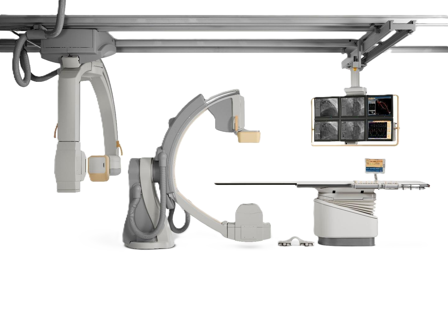
Philips FD10

G-Stand Cath Lab System

Developed through partnerships with the world's busiest cath labs, the FD10 delivers superb image quality at a low X-ray dose — with instant multi-modality access and advanced, easy-to-use image processing tools.

Key Performance Figures

100kW
Generator Power
25cm
Max Field of View (diag.)
75%
DQE at 0 lp/mm
85kW
Max Tube Loadability

Green Product — saves at least 12% energy vs. prior systems. 83% of lead content eliminated through counterbalance design.

Generator Velara 100 kW
X-ray Tube MRC-GS 0508
Stand Type G-stand (Ceiling / Floor)
Patient Weight Limit 250 kg
Image Storage 100,000 images
Green Certified Yes — asimpleswitch.com
System Overview

The G-shaped stand — built for cardiologists, by cardiologists

The FD10's unique G-stand reaches the groin without repositioning and covers a wide range of projections. Paired with the MRC-GS 0508 tube and next-generation flat detector, it delivers the speed and clarity that complex interventions demand.

G-Shaped Gantry

Maximises speed and patient access. Ceiling version: 260 cm motorised longitudinal travel at 15 cm/sec with auto-stops. BodyGuard capacitive patient protection at up to 25°/sec.

Xper Cardiovascular Table

Free-floating carbon-fibre tabletop, 319 × 50 cm. Supports up to 250 kg; CPR-compatible at any position. Optional tilt, cradle, and OR-compatible table series.

DoseWise Philosophy

Smart Beam management, automatic exposure controls, and real-time dose awareness — three strategies to protect patients and staff while maintaining image quality.

Xper User Interface

Fully customisable per user group and application. Complete system control available at tableside. Xper stands for X-ray Personalized.

DICOM Integration

Fast Ethernet DICOM Image Interface. Exports in XA Multi Frame or Secondary Capture formats to PACS, Xcelera, and HIS/RIS systems with continuous autopush option.

CX50 Ultrasound Integration

Philips CX50 ultrasound can be fully integrated via a one-click connection. Patient data shared automatically — no workflow duplication, controlled from tableside.

Imaging & Dose Management

DoseWise — a philosophy
that protects everyone

The FD10 integrates dose reduction at every level — from filtration hardware to real-time staff monitoring — without compromising image quality.

DoseWise

A set of techniques, programmes, and practices that ensures excellent image quality while protecting people in X-ray environments. Three highly effective strategies.

Smart Beam Management

Removes unwanted soft radiation and minimises scatter through SpectraBeam filtration.

Less Time

Automatic exposure controls to maximise dose efficiency throughout the procedure.

More Awareness

Clear, real-time dose information on screen so you always choose the right balance.

Dynamic Flat Detector

37 cm (14") housing, 28 cm physical detector, 25 cm maximum field of view. 1024 × 1024 px at 14-bit depth. Zoom fields at 25, 20, and 15 cm diagonal.

DoseAware (Optional)

Personal Dose Meters (PDMs) wirelessly feed real-time radiation exposure data to a basestation LCD — so staff can immediately see and adjust their behaviour.

Fluoroscopy Modes

Three programmable fluoro modes per application. Default storage of the last 10 seconds, expandable to 999 images. SpectraBeam, SPIRIT filters, and Xres algorithm.

Roadmap Pro

Advanced subtraction angiography roadmapping with automatic Motion Compensation — compensates for movement artefacts that could conceal clinical information.

Digital Acquisition

Virtually unlimited acquisition programmes for DSA and DA. Up to 1024 × 1024 pixels; standard storage 100,000 images. Frame rates 3.75, 7.5, 15, and 30 fps.

XperSwing & Rotational Scan

G-stand dual-axis swing in 3D arc for coronary imaging. Rotational scan at 55°/sec (head) and 30°/sec (side). Feeds 3D-CA and 3D-RA reconstruction tools.

01 G-Stand Design

Built for Cardiologists, by Cardiologists

The FD10's unique G-shaped stand architecture reaches the patient's groin without table repositioning and covers a wide range of projections — engineered through partnerships with the world's busiest cath labs to eliminate workflow bottlenecks.

  • 260 cm motorised longitudinal travel at 15 cm/sec
  • Rotation 120° LAO / 120° RAO up to 25°/sec in head position
  • BodyGuard capacitive patient protection
  • Programmable auto-stops: park, cardio, neuro, peripheral
Built for Cardiologists, by Cardiologists
02 DoseWise Philosophy

Three Strategies, One Goal — Less Dose

DoseWise is not a feature — it is a philosophy. The FD10 integrates dose management at every system level, combining hardware filtration, automatic exposure controls, and real-time staff monitoring to protect everyone in the room.

  • SpectraBeam filtration removes low-energy scatter radiation
  • DoseAware wireless PDMs for real-time staff monitoring
  • Automatic exposure controls minimise procedure time
  • Clear on-screen dose display throughout the procedure
Three Strategies, One Goal — Less Dose
03 Clinical Integration

One Workspace, Every Modality

The FD10 connects natively to the Xcelera clinical platform, consolidating X-ray, ultrasound, CT, MR, nuclear medicine, ECG, and electrophysiology data in a single workspace — so your team spends less time navigating systems and more time treating patients.

  • One-click CX50 ultrasound integration from tableside
  • DICOM XA Multi Frame and Secondary Capture export
  • Continuous autopush to PACS, Xcelera, HIS/RIS
  • OR-compatible Xper table series available
One Workspace, Every Modality
Technical Specifications

Detailed system parameters

Iso-centre to Floor 106.5 cm (ceiling and floor)
Longitudinal Movement (ceiling) 260 cm motorised + manual at 15 cm/sec — auto stops at park, cardio, neuro, peripheral
G-stand Rotation (head, ceiling) 120° LAO, 120° RAO up to 25°/sec
G-stand Rotation (side, ceiling) 45° LAO, 45° RAO up to 18°/sec
G-stand Angulation (head) 45° cranial, 45° caudal up to 18°/sec
G-stand Angulation (side, ceiling) 120° cranial, 120° caudal up to 25°/sec
Focal Spot to Iso-centre Up to 76.5 cm
Source Image Distance 86.5 – 123 cm
G-stand Depth 105 cm
Programmable Positions Standard 2; up to 10 with optional APC
Patient Protection BodyGuard capacitive sensing
Detector Housing Size 37 cm (14") diagonal, including BodyGuard
Physical Detector Size 28 cm (11") diagonal
Maximum Field of View 25 cm (10") diagonal square
Image Matrix 1024 × 1024 pixels at 14-bit depth
Detector Zoom Fields 25, 20, and 15 cm diagonal square formats
Pixel Pitch 184 × 184 µm
Nyquist Frequency 2.72 lp/mm
DQE at 0 lp/mm 75%
MTF at 1 lp/mm > 60%
Digital Output 1k² and 512² at 8 or 10-bit depth
Generator Type Velara — microprocessor-controlled 100 kW high-frequency converter
Voltage Range 40 – 125 kV
Maximum Current 1,250 mA at 80 kV
Nominal Power 100 kW (1,000 mA at 100 kV)
Minimum Switching Time 1 ms (quartz-controlled)
X-ray Tube MRC-GS 0508
Focal Spot / Loadability 0.5 / 0.8 — max 45 kW / 85 kW
Anode Heat Dissipation 3,200 W
Max Anode Cooling Rate 910 kHU/min
Fluoro Power (10 min) 4,500 W
Pre-filtration SpectraBeam: 0.2, 0.5, 1.0 mm Cu equivalent
Cooling Direct anode oil cooling, 200 mm anode diameter
Tabletop Material Radio-translucent carbon fibre
Tabletop Dimensions 319 × 50 cm
Height Adjustment (motorised) 79 – 104 cm
Metal-Free Overhang 125 cm
Longitudinal Float 120 cm
Transversal Float 36 cm
Max Patient Weight 250 kg + 500 N additional (CPR)
Optional Tilt Range ±17° (head-down / head-up) at 2°/sec
Optional Cradle Range ±15° at 3°/sec
OR Compatibility Xper OR Table series available for OR environment
Interventional Tools

A platform for advanced
clinical applications

Philips continues to enhance the Xper family's interventional capabilities — from 3D coronary reconstruction to electrophysiology navigation and whole-body vascular imaging.

Xcelera Integration: One access point for all relevant information — X-ray, ultrasound, CT, MR, nuclear medicine, ECG, and electrophysiology. One workspace for documentation, viewing, quantification, and reporting.

StentBoost

Instant, enhanced visualisation of stent position and deployment while the catheter is still in place. StentBoost Subtract improves stent-vasculature relationship visualisation.

Allura 3D-CA

3D model of 2D coronary artery images. Provides optimal viewing angles of lesions and bifurcations, and TrueView map for stent length selection.

Allura 3D-RA

Extensive 3D vascular visualisation from a single rotational acquisition. Covers cerebral, abdominal, and peripheral vasculature. Fully table-side controlled.

CT TrueView

All 3D-CA benefits from a CT diagnostic image. CTO Navigator overlays 2D exposure run over segmented cardiac CT data — matched manually or automatically.

EP Navigator

Fluoroscopy overlay of a 3D heart model. Catheters and 3D anatomy shown in real time, with fully automatic heart segmentation and single-click left atrium visualisation.

EP Cockpit

Integrates EP equipment on a single moveable ceiling rack. Reduces staff and patient radiation by up to 80%. EP cockpit XL adds a 56-inch high-resolution display.

QCA / LVA / RVA

Coronary stenosis measurement, left and right ventricular analysis, vascular analysis, and ejection fraction. Full Autocal option for calibration without additional acquisitions.

Subtracted Bolus Chase

Hand-held speed controller for peripheral run-off studies (ceiling FD10 only). Eliminates repeat exposures through accurate speed-matched subtraction.

Ambient Experience

Dynamic lighting and sound personalisation to reduce patient stress, improve clinical workflow, and differentiate your facility from the competition.

whatsapp-icon Customer Support